目次
Kager fat padの動態に関する重要な知見
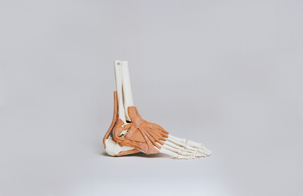
アキレス腱断裂後や足関節周囲外傷後に問題となることが多いのがKager fat padの癒着です.
Kager fat padの癒着によって足関節の背屈可動域が制限されることは多いですし,アキレス腱や下腿三頭筋の滑走性低下によって足関節底屈筋力の低下をきたす症例も少なくありません.
今回はKager fat padの動態に関する重要な知見を示した研究論文をご紹介させていただきます.
今回ご紹介する論文
Knee Surg Sports TraumatolArthrosc
. 2020 Jan;28(1):148-154. doi: 10.1007/s00167-019-05585-1. Epub 2019 Jun 29.
Pressure changes in the Kager fat pad at the extremes of ankle motion suggest a potential role in Achilles tendinopathy
F Malagelada 1 2, J Stephen 3 4, M Dalmau-Pastor 5 6 7, L Masci 8, M Yeh 4, J Vega 5 6 9 10, J Calder 3 4
Affiliations expand
PMID: 31256217 DOI: 10.1007/s00167-019-05585-1
今回ご紹介する論文は2020年に掲載された論文です.
研究の背景
Introduction: The Kager fat pad is one of the largest soft tissue structures local to the ankle joint, yet it is poorly understood. It has been hypothesised to have a role in Achilles tendinopathy. This study aimed to investigate the pressure areas in the Kager fat pad adjacent to the Achilles tendon and to assess the anatomy and deformation of the Kager fat pad in cadavers.
Kager fat padは足関節に存在する最も大きな軟部組織の1つですが,その実態は不明な点も多いです.
アキレス腱症に関与しているといった説もあります.
この研究ではアキレス腱に隣接するKager fat padの圧迫部位を調査し,死体でKager fat padの解剖学的構造と変形を評価することを目的としております.
研究の方法
Methods: Twelve fresh frozen cadaveric ankles (mean age 44 years, range 38-51) were mounted in a customized testing rig, enabling plantar flexion and dorsiflexion of the ankle, with the Achilles tendon loaded. A needle tipped pressure sensor was inserted in two areas of the Kager fat pad under ultrasound guidance (retrocalcaneal bursa and at 3 cm proximal from Achilles insertion). Pressure readings were recorded at different flexion angles. Following testing, the specimens were dissected to expose the Kager fat pad and retrieve it for analysis. MRI images were also taken from three healthy volunteers and the Kager fat pad deformation examined.
12例の新鮮凍結死体の足関節(平均年齢44歳,範囲38-51)をカスタマイズしたテストリグに装着し,アキレス腱に負荷をかけた状態で足関節底屈・背屈運動が可能となるようなセッティングを行いました.
超音波ガイド下で針先の圧力センサーをKager fat pad の2つの部位(踵骨後部の滑液包とアキレス腱挿入部から3cmの近位部)に挿入しております.
異なる背屈・底屈角度で圧力測定値を記録しております.
試験後に検体を解剖してKager fat pad を露出させ分析のために回収しております.
また3名の健康なボランティアからMRI画像を撮影しKager fat padの形態変化を調査しております.
研究の結果
Results: Mean pressures significantly increased in all specimens at terminal ankle plantar and dorsi flexion in both regions (p < 0.05). The Kager fat pad was consistently adherent to the Achilles at its posterior aspect for a mean length of 7.7 cm (SD 0.27, 89% of KFP length). The most distal part of the Kager fat pad was the exception and it detached from the Achilles to give way to the retroalcaneal bursa for a mean length of 0.92 cm (SD 0.24, 11% of KFP length). The bursal space is partially occupied by a constant ‘wedge’ extension of Kager fat pad. The mean volume of the whole Kager fat pad was 10.6 ml (SD 3.37). Video and MRI demonstrated that the Kager fat pad undergoes significant deformation during plantar flexion as it is displaced superiorly by the Achilles, with the wedge being forced into the retrocalcaneal bursal space.
平均圧力はすべての検体において,足関節背屈・底屈最終域で両部位ともに有意に上昇しております(p<0.05).
Kager fat padはその後面でアキレス腱に平均7.7cm(SD 0.27、KFP長の89%)の長さで一貫して密着しておりました.
しかしながらKager fat padの最遠位部分は動態が異なり,アキレス腱から独立して平均0.92cm(SD 0.24,KFPの長さの11%)で後踵骨滑液包へと移動しておりました.
滑液包の空間は部分的にKager fat padの一定の くさび の延長によって占められておりました.
Kager fat pad 全体の平均体積は10.6ml(SD 3.37)でありました.
ビデオとMRIによって足関節底屈背屈運動時にKager fat padがアキレス腱によって上方向に変位し,後踵骨滑液包に押し込まれることでKager fat pad の形が大きく変化することが示されました.
研究の結論
Conclusion: The Kager fat pad does not remain static during ankle range of motion, but deforms and its pressure also changes. This observation supports the theory that it acts as a shock-absorber to the Achilles tendon and pathological changes to the fat pad may be clinically important in the development of Achilles tendinopathy.
Kager fat padは足関節の可動域運動時に静止しているのではなく形態を変化させその圧力も変化することが明らかとなりました.
この観察結果はアキレス腱の衝撃吸収体として機能しているという理論を裏付けるものであり,Kager fat padの病的変化はアキレス腱症の発症において臨床的に重要であると考えられます.


今回はKager fat padの動態に関する重要な知見を示した研究論文をご紹介させていただきました.
これは非常に興味深い結果ですね.
遠位部とその他では動態が異なるといった点は臨床上も非常に重要なポイントになりそうですね.
今後さらなる研究が期待されますね.