目次
足関節背屈方向のストレッチングは足部回内位・回外位どちらで行った方がよい?
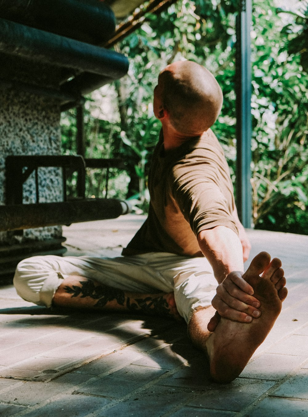
理学療法士・作業療法士が足関節背屈方向のストレッチングを実施することは多いと思います.
昔から何の意味もなく下腿三頭筋のストレッチングを行う理学療法士って多いですよね.
もはや癖みたいになっている場合も多いでしょう…
ただ足関節背屈方向のストレッチングを行い場合には足部を回内位・回外位のどちらで行うのが有効なのでしょうか?
もちろん腓腹筋の内側頭・外側頭どちらを伸張したいかといった目的によるところも大きいですが…
今回は足関節背屈方向のストレッチングは足部回内位・回外位どちらで行った方がよいかを明らかにした研究論文をご紹介させていただきます.
今回ご紹介する論文
Randomized Controlled Trial Foot Ankle Int
. 2014 Jan;35(1):63-70. doi: 10.1177/1071100713513433. Epub 2013 Nov 20.
The effect of subtalar joint position on dorsiflexion of the ankle/rearfoot versus midfoot/forefoot during gastrocnemius stretching
Marie A Johanson 1, Amy DeArment, Krystol Hines, Erin Riley, Meghan Martin, Justin Thomas, Kathleen Geist
Affiliations expand
PMID: 24259750 DOI: 10.1177/1071100713513433
今回ご紹介する論文は2014年に掲載された論文です.
研究の背景
Background: Limited ankle joint dorsiflexion passive range of motion (PROM) has been associated with common chronic lower extremity conditions, and clinicians often instruct patients in stretching exercises to increase dorsiflexion. However, little is known about how subtalar joint (STJ) position affects dorsiflexion at the midfoot/forefoot versus ankle/rearfoot during gastrocnemius stretching. The purpose of this study was to determine if more dorsiflexion occurs at the ankle/rearfoot and less at the midfoot/forefoot during gastrocnemius stretching with the STJ positioned in supination versus pronation.
足関節背屈の他動的可動域制限は,慢性下肢疾患と関連しており,臨床医はしばしばクライアントに背屈可動域の拡大を目的としてストレッチ運動を指導します.
しかしながら腓腹筋ストレッチ時の中足部・前足部と足関節・後足部の背屈に,距骨下関節(STJ)の肢位がどのように影響するかについてはほとんど知られておりません.
この研究では距骨踝関節(STJ)の肢位が回内位と回外位では,どちらが腓腹筋の伸張運動時にに足関節・後足部で背屈が生じやすく,どちらで中足部・前足部では背屈が生じにくいかを明らかにすることを目的としております.
研究の方法
Methods: In this repeated measures design, 27 participants (23 females, 4 males; mean age = 31.3 years, SD = 10.7) with current or recent history of lower extremity chronic conditions and less than 10 degrees ankle dorsiflexion measured with the knee in extension on the involved side(s) performed five 30-second gastrocnemius stretching trials in pronation and supination on each side in a randomly determined sequence. A 7-camera Vicon Motion Analysis System and an AMTI force plate were used to measure midfoot/forefoot dorsiflexion, ankle/rearfoot dorsiflexion, knee extension, and normalized vertical ground reaction force.
この研究では下肢慢性疾患の既往歴または最近の病歴があり,膝関節伸展位での足関節背屈可動域が10°未満の27例(女性23人,男性4人,平均年齢=31.3歳,SD=10.7)を無作為に決められた順序で,片側ずつ30秒間の腓腹筋ストレッチを回内位と回外位で5回ずつ行っております.
7台のカメラを備えたVicon Motion Analysis SystemとAMTI社のフォースプレートを用いて,中足部/前足部の背屈可動域,足関節/後足部の背屈可動域,膝関節伸展可動域,正規化された垂直方向の地面反力を測定しております.
研究の結果
Results: Two-way repeated measures ANOVA revealed a significant increase in midfoot/forefoot dorsiflexion when stretching in pronation compared to supination (P < .001). ANOVAs also demonstrated significantly more extension of the knee when stretching in supination compared to pronation (P < .001), and increased normalized vertical ground reaction force when stretching in supination compared to pronation (P = .032). With the numbers available, no significant difference in ankle/rearfoot dorsiflexion when stretching in supination compared to pronation could be detected (P > .05).
二元配置反復測定分散分析の結果,中足部および前足部の背屈可動域は,回外位に比較して回内位でストレッチしたときに有意に増加しました(P < 0.001).
また分散分析では,回内位に比べて回外位でストレッチしたときに,膝関節伸展有意に増加し(P < 0.001),回内位に比べて回外位でストレッチしたときに,正規化された垂直方向の地面反力が増加しました(P = 0.032).
足関節・後足部の背屈可動域については,有意差は見られませんでした(P > 0.05).
研究の結論
Conclusion: Gastrocnemius stretching in pronation resulted in more dorsiflexion at the midfoot/forefoot than stretching in supination.
腓腹筋を回内位でストレッチすると,中足部/前足部の背屈可動域が回外位でストレッチするよりも大きくなることが明らかとなりました.


今回は足関節背屈方向のストレッチングは足部回内位・回外位どちらで行った方がよいかを明らかにした研究論文をご紹介させていただきました.
解剖学的な視点で考えれば当然の結果な気がしますが,中足部・前足部の背屈可動域の拡大を図りたい場合には足部を回内位として可動域運動を行うのがよさそうですね.